Microsatellite Instability (MSI) Analysis Comparing Normal and Tumor Samples
MSI analysis is essential for tumor research, such as colorectal, triple negative breast, EMAST (Elevated Microsatellite instability at Selected Tetranucleotide repeats) and bladder cancer. This instability is due to either insertion or deletion of repeating units during DNA replication and failure of the mismatch repair system (MMR) to correct these errors.
The MSI Application in GeneMarker® software is directly linked to the main analysis screen and is an excellent tool for analysis from kits such as the Promega MSI Analysis System, or Thermo Fisher TrueMark™ MSI Assay separated with ABI PRISM® , SeqStudio™ or Spectrum Compact CE Systems genetic analyzers. GeneMarker software's MSI Analysis module compares tumor to normal samples based on peak-to-peak comparison to differentiate between MSH-H and MSS samples. See figures 1 and 2 below for an overview of the MSI application.
MSI Normal and Tumor Electropherogram Overlay View
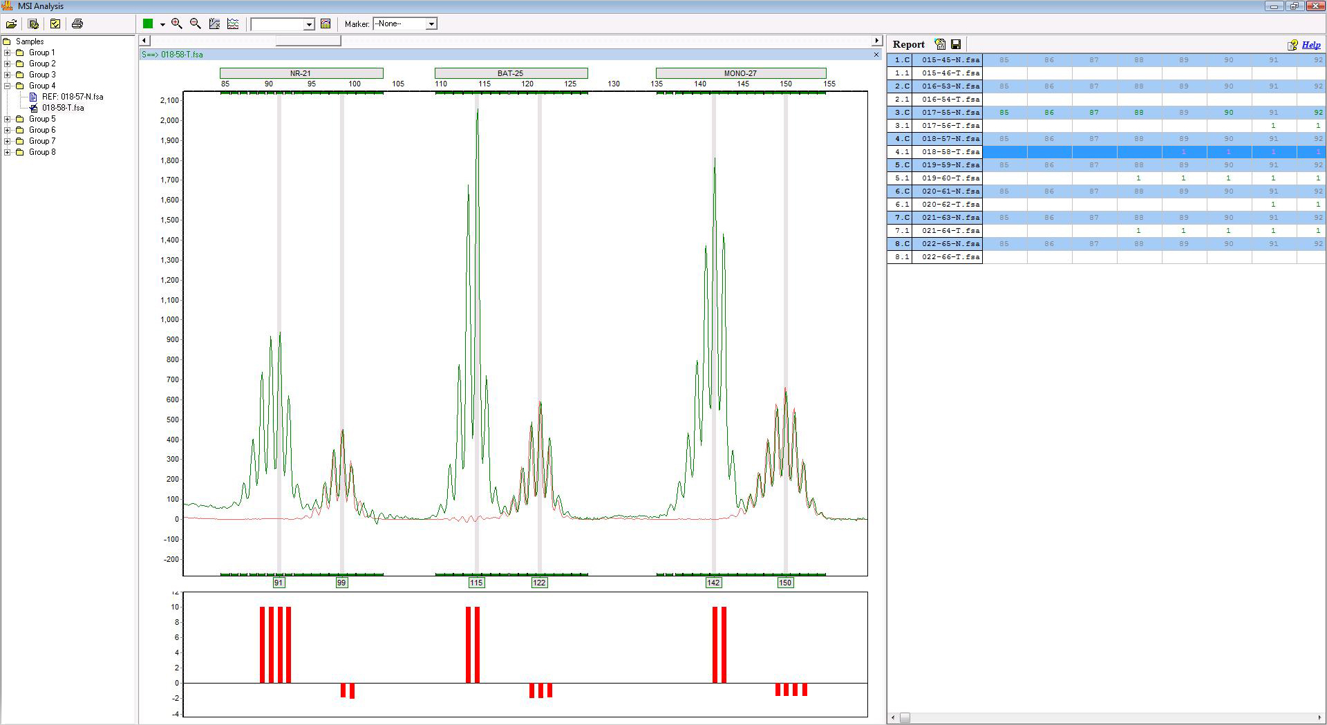
Figure 1: The tumor electropherogram for each sample is displayed in an overlay with the normal electropherogram (red trace) for easy visualization of any instability.
Customized Reporting
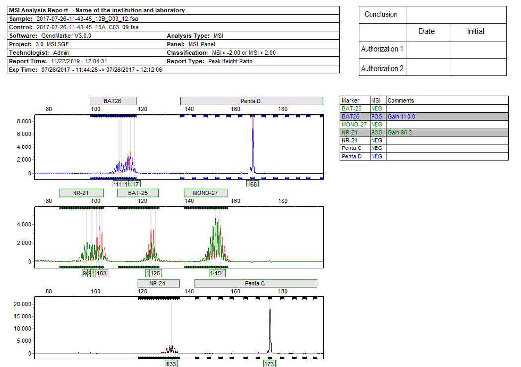
Figure 2: Includes a header for electronic signature, trace overlay (tumor and normal), trace comparison histogram and gain/loss table.
Application Notes:
MLPA® is the registered trademark of MRC Holland